What Are the Signs and Symptoms of ILC?
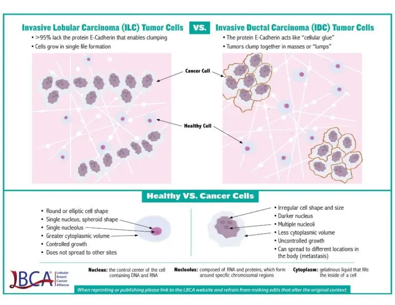
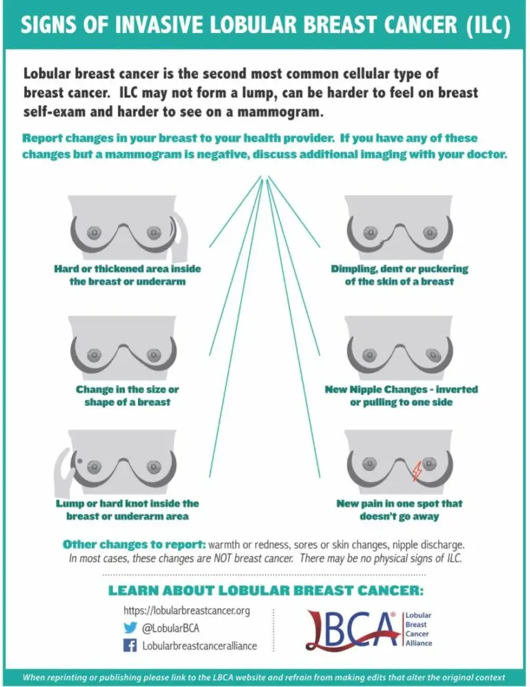
Because ILC usually does not form lumps, it is much harder to detect with screening, advanced imaging, and self-exams. Symptoms of ILC tumors can range from none to changes in the appearance of the nipple or breast, such as dimpling, hardening of the breast, swelling, or pain. On self-examination, ILC can sometimes be felt as a firmness or mass.
How is ILC currently treated?
Lobular breast cancer is currently treated like the more common IDC/NST despite its differences. An individual’s treatment plan for ILC depends on many factors, including the size and grade of the cancer, genetic factors, lymph node involvement, and the patient’s overall health and individual preferences. At present, there are no ILC-specific treatment guidelines. The standard of care for treatment of all stages of hormone receptor positive ILC is the same as treatment of hormone receptor positive IDC/NST. More research is needed to identify ILC-specific treatment protocols.
Hormone therapy is recommended for both pre- and post menopausal patients with hormone receptor positive ILC. Aromatase inhibitors and tamoxifen are used most commonly. Chemotherapy may be beneficial for some patients with ILC, but there is ongoing research into when it is most effective for those with early stage hormone receptor positive disease.
What Are the Specific Challenges of Metastatic ILC?
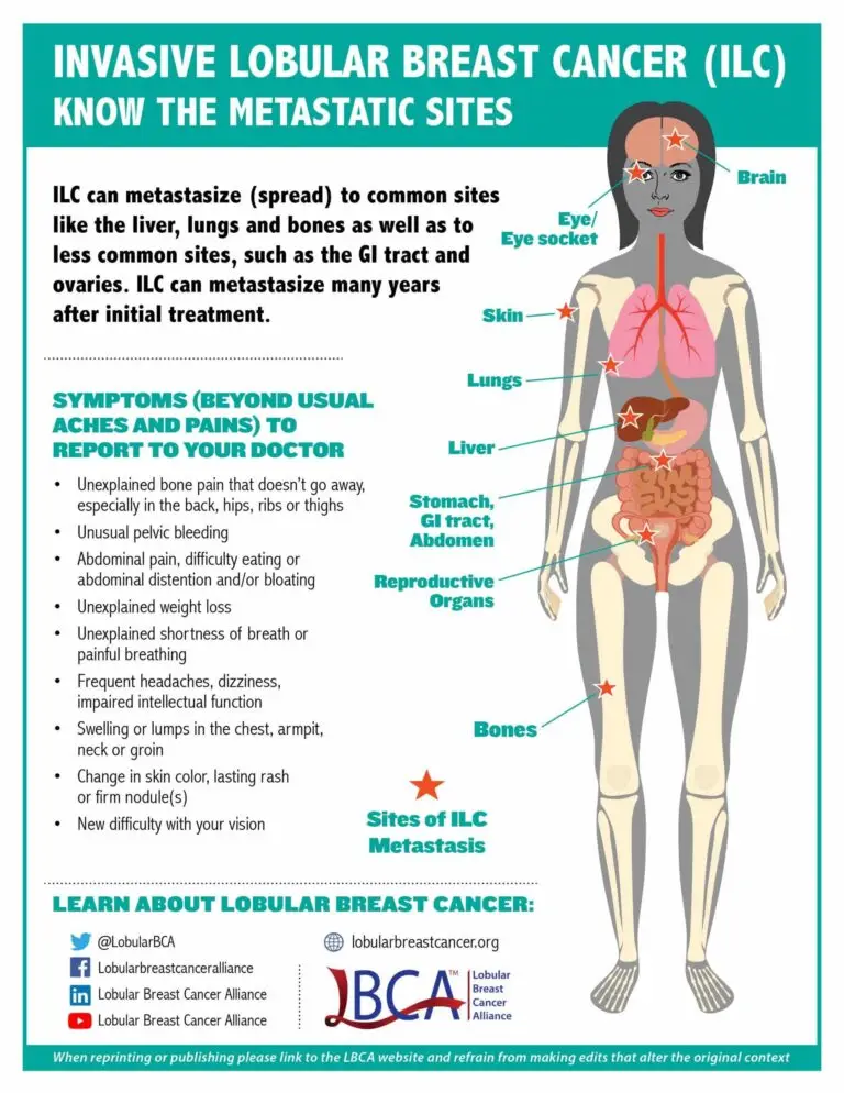
Lobular breast cancer can metastasize to unusual places. Similar to IDC/NST, ILC can metastasize to the bones, brain, liver, and lungs. However, ILC can also spread to unique sites such as the gastrointestinal tract (stomach, small intestine, and colon), gynecological organs (ovaries, uterus), the peritoneum (abdominal lining), and in rarer cases leptomeninges (lining of the brain and spinal cord) and orbital tissues (tissues around the eye).
Lobular breast tumors frequently recur many years after primary diagnosis, and are harder to monitor when metastatic.
While lobular breast cancer, like IDC/NST, can recur any time after initial diagnosis, studies show that ILC often recurs later than IDC, more than 10 years after the initial diagnosis of cancer. Since ILC typically grows in a linear pattern through the breast without distorting the surrounding structures or forming a lump, ILC is difficult to detect on mammography and ultrasound or to visualize when metastatic. The linear growth pattern of ILC sometimes makes it difficult to detect metastatic ILC with current imaging technology such as traditional computed tomography (CT), positron emission tomography (PET) and bone scans and not all metastatic lobular sites may be seen. Sometimes tests such as colonoscopy/endoscopy or biopsies are necessary to confirm metastatic ILC disease. FES PET scans may also be useful in detecting or monitoring metastatic ILC.
Because of the potential for ILC to metastasize to uncommon sites, it is important for people with lobular breast cancer and their oncologists to be aware of these differences, and to discuss the importance of recognizing and reporting possible symptoms referring to these unusual sites of metastases.
Additional Information on Lobular Breast Cancer

ILC Overview Brochure for Download
This in-depth brochure includes an overview of ILC, its key features, how it differs from other types of breast cancer, as well as current information on imaging. It also contains a list of publications and the resources LBCA provides. The brochure is available in both a digital version to share via email or on social media, and as a download to print.
Information and Resources for Patients
To stay up to date on ILC research news and how you can get involved to help raise awareness, sign up for the LBCA newsletter.